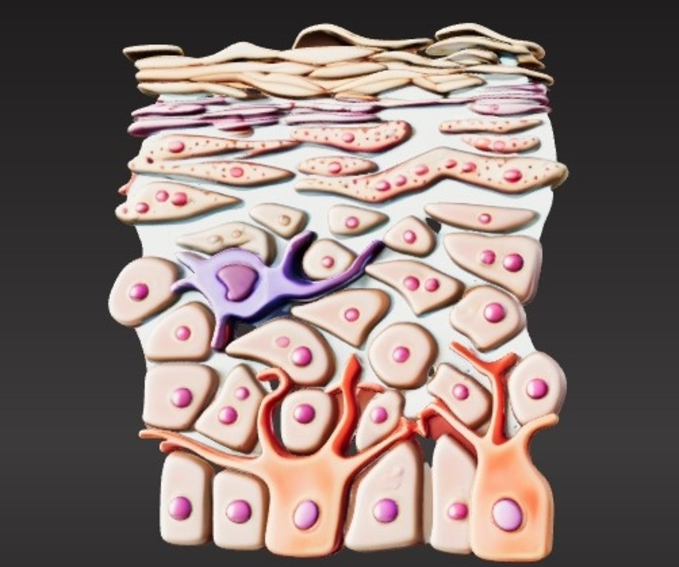


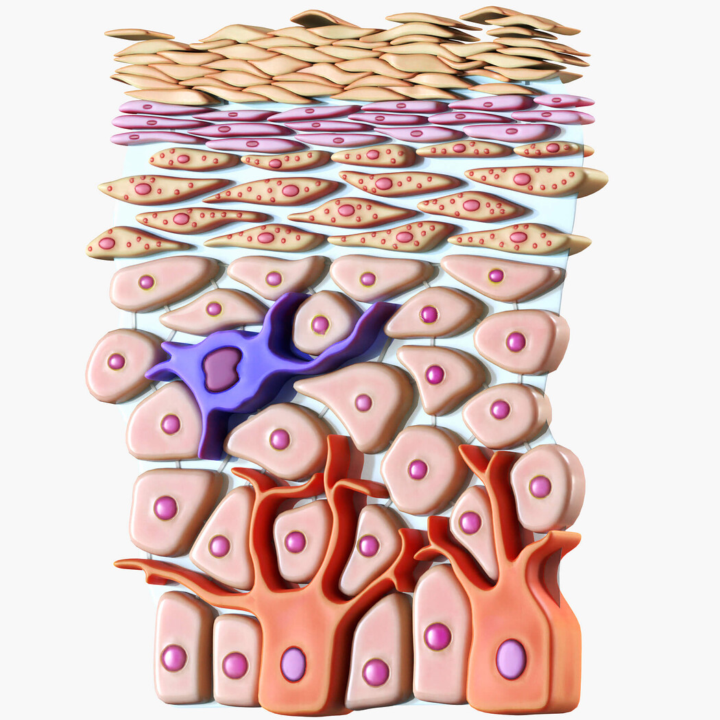


3D Model of Epidermis Cross Section
This 3D model of the epidermis cross-section is an anatomically accurate representation of the skin's outermost layer, highlighting its intricate structure and organization. Designed for educational, medical, and artistic purposes, it provides a detailed view of the layers and cellular components of the epidermis.
Key features:
Displays all layers of the epidermis: stratum corneum, stratum lucidum (if applicable), stratum granulosum, stratum spinosum, and stratum basale. Includes keratinocytes, melanocytes, Langerhans cells, and Merkel cells, accurately depicted within their respective layers. Highlights the basement membrane and its connection to the dermis. Optional enhanced views of skin appendages such as hair follicles, sweat glands, and sebaceous glands integrated into the model. Realistic textures and proportions to mimic the microscopic appearance of the epidermis. Applications:
Medical education for teaching skin anatomy, structure, and function. Research in dermatology, including studies on skin conditions, wound healing, and regeneration. Artistic reference for creating detailed visualizations of skin anatomy. The model is optimized for 3D printing and digital exploration, provided in widely compatible file formats such as STL and OBJ. This epidermis cross-section model is a valuable resource for educators, researchers, and artists seeking a precise and clear representation of this vital part of human anatomy.
Model Details
- FormatsPLY, FBX, STL, OBJ, BLEND, DAE
- Polygons499976
- Vertices267540
- animatedno
- materialsyes
- texturesyes
- riggedno
- uvsyes
- 3d print readyyes
You will get 9 files
All files previously purchased will always be available for download in your Library